
11 April 2019
With the aim of developing safer nanoparticles for biomedical applications, scientists of the group Nano-Cell Interactions at INM – Leibniz-Institute for New Materials and Pharmacelsus GmbH in Saarbrücken have studied a three-dimensional liver cell model. The model allows analysis of biomarkers in combination with information on penetration depth of the particles into the tissue. Therefore it is better suited for nanoparticle testing and development than conventional two-dimensional cell models, where cells are growing as a single layer.
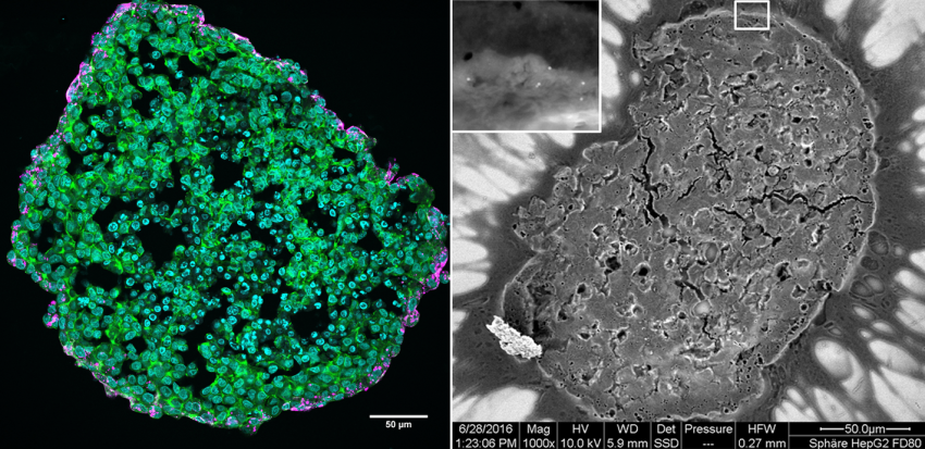
Left: Fluoreszence image of liver cell micro tissue (green: cytoskeleton, blau: nuclei) with silica nanoparticles (magenta) located at the border of the micro tissue.
Right: Elektron microscopy image of a liver cell drop. Individual nanoparticles are visible in the outer rim of the cell drop (white in detail on top left).
Artificial nanoparticles provide novel possibilities for biomedical applications. It is important to identify possible risks early in the developmental process. As many drugs and metabolites are catabolized in the liver, the organ plays an important role for testing new biomedical products. ″Nanoparticle-based contrast agents are injected directly into the blood and are eventually transported to the liver. For this reason we want to use advanced cell culture models of the liver for our savety assessments″, says Annette Kraegeloh, leader of the research group Nano-Cell Interactions at INM.
Classic 2D cell culture models behave differently than their 3D counterparts. „If we cultivate the liver cells as so called micro-tissues – small spheres with a diameter of about half a millimeter – they produce a protein that covers bile ducts in the liver. Moreover, the gene expression for enzymes that metabolize drugs is increased. They are therefore better suited as a model for the organ than the 2D model, where only one layer of liver cells grows on a plastic surface“, explains Jana Fleddermann, first author of the now published study. The 3D model allows the researchers to determine how deep the nanoparticles are penetrating into the tissue – another advantage of the micro tissue model compared to commonly used 2D models.
The scientists at INM and Pharmacelsus GmbH tested the micro tissue model with silica particles (SiO2) with a diameter of 100 nm. Silica nanoparticles are a promising material for applications in drug delivery, biosensing and as carrier for contrast agents in medical imaging. The particles were loaded with a fluorescing dye in order to make them visible under the microscope. The maximum penetration depth measured was 20 µm (about 3 cell layers). The silica particles did not harm the cells and did not interfere with tissue growth and cell behavior in a perceivable way. To further improve safety assessment of nanoparticles, different cell types that occur in the liver should be grown together to develop an even more realistic cell culture model.